
EGD Techniques and Their Applications in Diagnosis
An EGD (esophagogastroduodenoscopy) is a minimally invasive endoscopic procedure. It examines the esophagus, stomach, and duodenum, and physicians use it to diagnose conditions such as ulcers and gastrointestinal cancers. Various techniques are employed during the procedure, each of which contributes to a more accurate diagnosis. Here is more information on the procedure, the reasons it is performed, and the techniques used:
What Is an EGD?
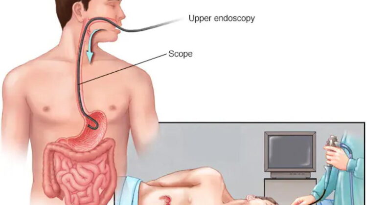
An EGD is a diagnostic endoscopic procedure that allows a physician to examine the upper gastrointestinal (GI) tract. A thin, flexible tube called an endoscope is passed through the mouth, and it captures real-time images of the GI lining using a fitted light and camera. These images help physicians assess tissue condition in detail. The procedure is performed under sedation, which helps keep the patient comfortable while allowing the physician to conduct a thorough examination. The upper GI tract regions examined during an EGD include:
- Esophagus: The muscular tube connecting the throat to the stomach
- Stomach: The organ responsible for breaking down food
- Duodenum: The first section of the small intestine
Why Is It Conducted?
Physicians recommend an EGD when a patient presents with symptoms suggesting a disorder of the upper GI tract; these symptoms may include persistent heartburn, difficulty swallowing, unexplained weight loss, abdominal pain, or gastrointestinal bleeding. The procedure can also be used to monitor known conditions. When other imaging studies identify abnormalities, an EGD provides a more detailed internal assessment, making it a versatile diagnostic tool.
An EGD also serves a therapeutic function in select cases. Physicians may use the endoscope to remove polyps or treat active bleeding sites, which makes the procedure both diagnostic and interventional. When a narrowed section of the esophagus is identified, physicians can dilate it during the same procedure. This eliminates the need for a separate intervention.
What Are the Available Techniques?
Several techniques are used during an EGD, which depends on the clinical objective. Esophageal dilation is a technique used to widen a narrowed section of the esophagus, and during the procedure, a physician passes a dilating instrument through the endoscope to stretch the affected area. Tissue sampling, or biopsy, is another standard technique. The physician passes a small instrument through the endoscope’s working channel to collect tissue samples for laboratory analysis.
How Does It Facilitate Diagnosis?
The EGD provides direct visualization of the upper GI tract, which makes it a more precise diagnostic tool than imaging studies like X-rays or CT scans. Physicians document findings in real time. When the camera reveals inflammation, lesions, or structural irregularities, the physician can record findings immediately, and biopsied tissue offers an additional diagnostic layer. Laboratory analysis can confirm infections, identify precancerous changes, or diagnose conditions like celiac disease.
Learn More From a Specialist
An EGD is a procedure that provides reliable diagnostic information about the upper gastrointestinal tract. Physicians use a range of techniques to identify and characterize conditions that affect the esophagus, stomach, and duodenum, and each technique adds value to the diagnostic process. If you have been referred for an EGD, consult your gastroenterologist to discuss which techniques may be used.